Learning Strategies for Brain Tumor Segmentation.
Accurate segmentation of brain tumor sub-regions is essential in the quantification of lesion burden, providing insight into the functional outcome of patients. In this regard, 3D multi-parametric magnetic resonance imaging (3D mpMRI)
this Project contain the implementation of paper we are working with the team from Jorden Univesity of science and technology , in this work we present a new based model on Neasted 3DUnet++ combined with attention mechanism Block for more effiency feature extracttion
the problem case study in 3D Biomedical image processing
- subject
Accurate segmentation of brain tumor sub-regions is essential in the quantification of lesion burden, providing insight into the functional outcome of patients. In this regard, 3D multi-parametric magnetic resonance imaging (3D mpMRI) is widely used for non-invasive visualization and analysis of brain tumors. Different MRI sequences (such as T1, T1ce, T2, and FLAIR) are often used to provide complementary information about different brain tumor sub-regions
- Problem
in many cases for processing Medical images to get better understanding of disease and impact on human being life such Brain tumor is most area for reseachers to improve system diagnosis in partuclar Task Segementation , last few years lunch of challenges BRATS for segmentation Brain tumor Sub-regrion many of studying came up to improve CAD system ,
- Solution
For automatic segmentation we will use Unet3d To predict the 3D Volumitric Shape of tumor: we used Neasted 3DUnet++ combined with attention mechanism Block for more effiency feature extracttion and we trained on indenpendently 3DUnet++ and 3DUnet++ combined with attention mechanism
introduction
Imaging Data Description
All BraTS multimodal scans are available as NIfTI files (.nii.gz) and describe a) native (T1) and b) post-contrast T1-weighted (T1Gd), c) T2-weighted (T2), and d) T2 Fluid Attenuated Inversion Recovery (T2-FLAIR) volumes, and were acquired with different clinical protocols and various scanners from multiple (n=19) institutions, mentioned as data contributors here.
All the imaging datasets have been segmented manually, by one to four raters, following the same annotation protocol, and their annotations were approved by experienced neuro-radiologists. Annotations comprise the GD-enhancing tumor (ET — label 4), the peritumoral edema (ED — label 2), and the necrotic and non-enhancing tumor core (NCR/NET — label 1), as described both in the BraTS 2012-2013 TMI paper and in the latest BraTS summarizing paper. The provided data are distributed after their pre-processing, i.e., co-registered to the same anatomical template, interpolated to the same resolution (1 mm^3) and skull-stripped.
environment-project
- setup the enviroment;
- install script shell
here you will need to run script shell to install all the dependencies needed for
run code :
- create kaggle account to access to the data API
- add path kaggle.json to script shell $path_api
-
create the enviromenet here you will need to run
conda create --name Segemnetation python=3.6 -
make sure the requirements.txt exist to the repo install the packges if you want fisrt neeed to run
pip install -r requirements.txt -
here you will need to run script shell to install all the dependencies needed automated setup whole project
chmod +x download_dataset.sh && ./automate_downlaod_data.sh ### run-project
- install script shell
here you will need to run script shell to install all the dependencies needed for
run code :
-
PostProcessig dataset Brast2020;
this tool built based on top of BET algorithm that publish from FSL and N4baisCorrection we automated the process and handle the data in 3D shape link Project Repository visited and run this First Stage
Model-Description
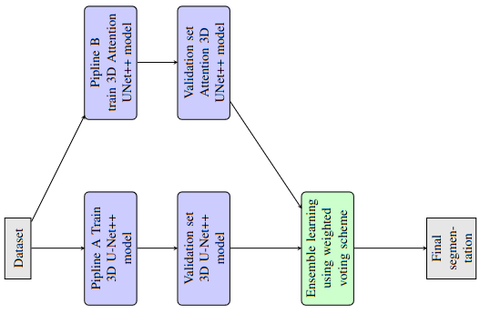
our Pipline divde into tow Stage to train model Unet++ and Unet++ with Attention Gate
-
The Full Pipline Ensemble learning:
in our Purposal we trained the model indenpendently and we combined the prediction both of the wieghts Checkpoints we provide open to downlaod following line Drive
Models Parameters Size MB 3D Attention Unet++ 30 M 101.5 MB 3D Unet++ 31 M 785 MB Stratgy used is Wieght Voting algorithm here’s the full picture
-
Ruuning the model indenpendently:
we provide the Yaml file configuration for Fine-Tuning the model brast2020.yml , has all the Hyper-Parameters to setup , we can change the model by updating the Model in config file which are [UNET3DPPATTEN ,UNET3DPP ] for run the model to train on GPU follwing the command :
python train.py --stage train --config brast2020.yaml --fold 0 -
Evaluation the model indenpendently:
we provide here also the evaluation model indenpendently , we can change the model by updating the Model in config file evaluate.yaml which are [UNET3DPPATTEN ,UNET3DPP ] for run the model to train on GPU follwing the command :
python predict.py --stage evaluate --config evaluate.yaml
Results
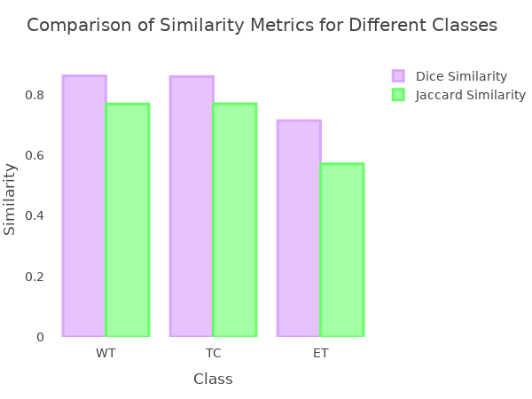
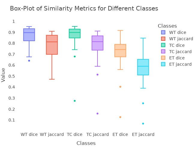
the performence of the both model we trained on indenpendently for 250 Epochs shows in the table and Diagram blow :
Table 1: show the Evaluation Metrics we used DCS and JSC
| Models | Dice Similarity WT | Dice Similarity TC | Dice Similarity ET | Jaccard Similarity WT | Jaccard Similarity TC | Jaccard Similarity ET |
|---|---|---|---|---|---|---|
| Pipline A | 0.88 | 0.87 | 0.73 | 0.79 | 0.78 | 0.59 |
| Pipline B | 0.82 | 0.82 | 0.69 | 0.72 | 0.72 | 0.54 |
| Ensemble Learning | 0.86 | 0.86 | 0.71 | 0.77 | 0.77 | 0.57 |
Figure : the virtualization of Results Bar Plot both of UNET3DPPATTEN ,UNET3DPP
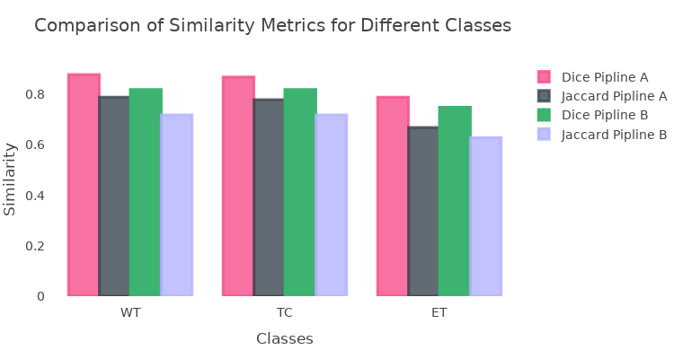
Figure : Results of Ensemble Wieght Voting Bar Plot both of Ensemble Learning
Prediction
we have a full Notebook has all the Quantative results used both modesl we trained on with Ensemble Learning results-attention-3d-unetpp-torchio
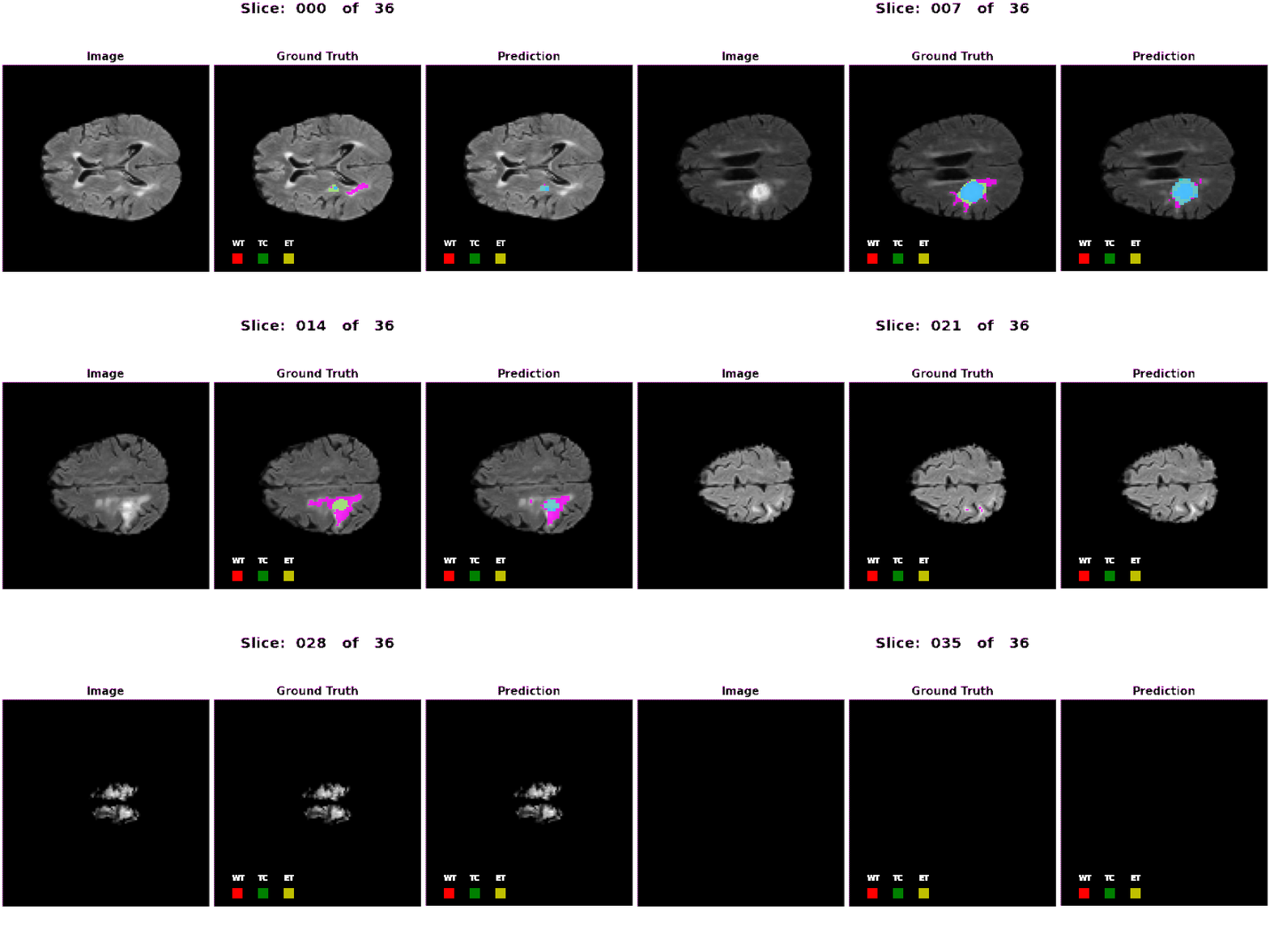
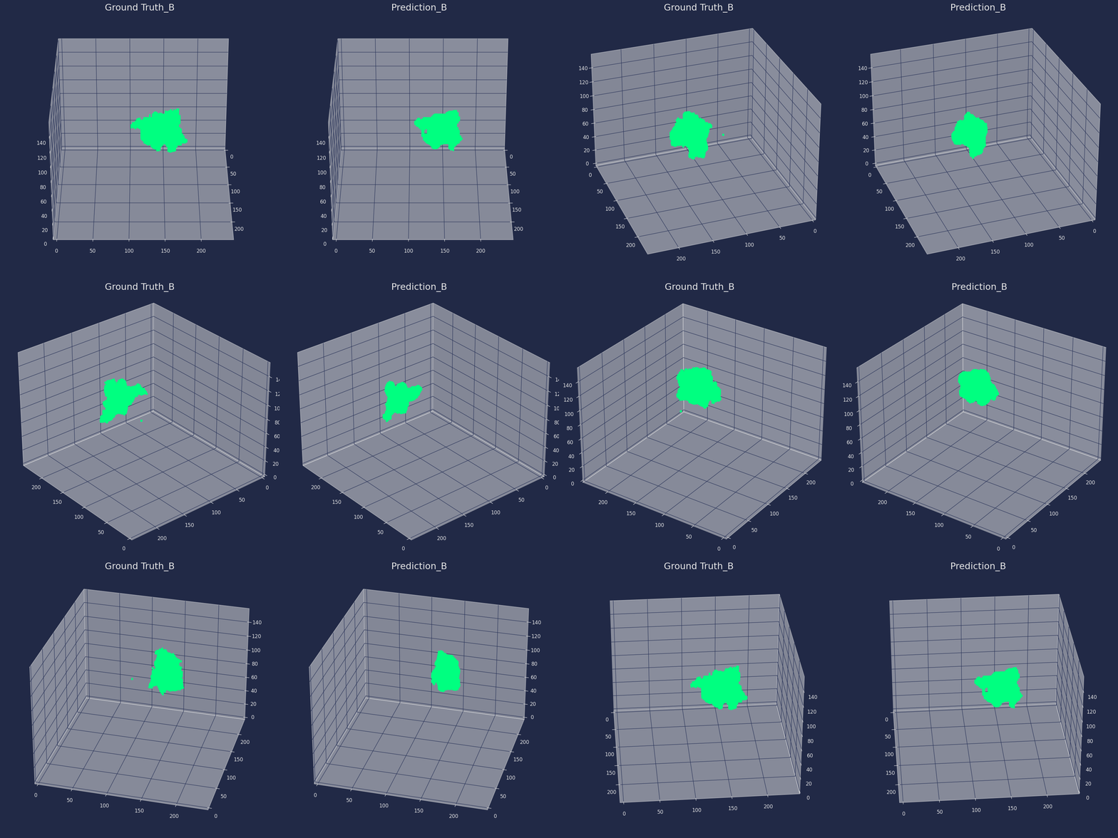
here the 3D prediction of Enesmeble Learning model both of Animation Slicer Frames and 3D Extraction Volumitric Data
- Animations Frames:
